


News
Takata Authors New Publication on Identifying Aberrant Muscles in the Carpal Tunnel
October 30, 2018
Congratulations to MSOP’s PhD student Sandy Takata on her newest publication in the Journal of Diagnostic Medical Sonography that describes how sonography can assist in identifying long flexor and lumbrical muscle bellies within the carpal tunnel. Proper identification of these structures and evaluation their relative impact on the median nerve may be important to understanding nerve compression or entrapment for prevention and treatment of carpal tunnel syndrome. Read the full text ⟩
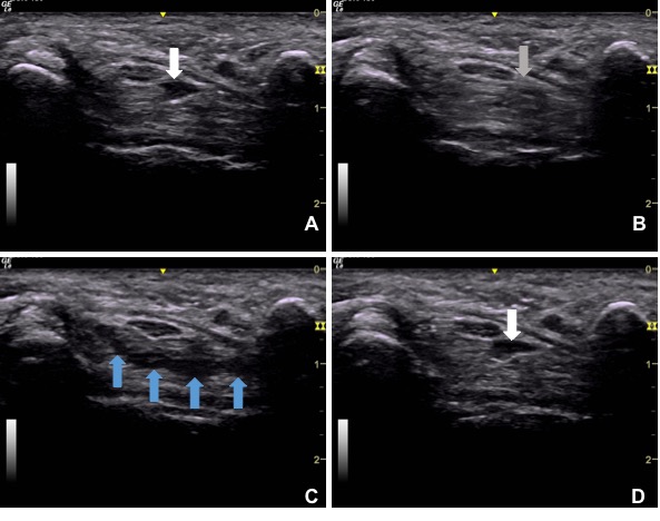
Image Above: Serial sonographic images of the left carpal tunnel at the level of the pisiform. The participant was asked to open her hand with extended fingers (A), slowly flex her fingers to make a fist (B), hold the fist position for one second (C), and extend the fingers once again (D). As the participant makes a fist, the long flexor muscle belly (white arrow in A) retracts proximally into the forearm and is replaced by the flexor tendon (gray arrow in B). When the fingers are fully flexed, the lumbricals enter the carpal tunnel (blue arrows in C). Once the fingers return to extension, the long flexor muscle belly is seen in the tunnel once again (white arrow in D).