Completed Research

Compression of the median nerve (asterisks) while holding a dental scaling instrument
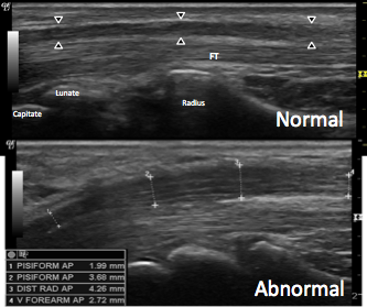
Together with researchers in the Laboratory for Investigatory Imaging at The Ohio State University, our research teams combine expertise in diagnostic sonography with clinical evaluation and treatment of musculoskeletal disorders to develop and evaluate novel, cutting-edge methods. Collaborative research projects have evaluated the utility of quantitative & qualitative grey-scale, Doppler, and contrast-enhanced techniques to investigate these novel methods and techniques for diagnosis (OSU research) and intervention (USC research).
The two programs have obtained grant funding, published numerous manuscripts, and given several national and international presentations.
Funding
Investigation into Staging CTS Using Portable Ultrasound and Doppler Imaging
R21-OH009907 (PI: Evans, Co-I: Roll)
National Institute for Occupational Safety and Health / Centers for Disease Control
Total Funding: $265,834
Funding Period: 9/1/2010 – 8/31/2012
Publications
Roll, S. C., Volz, K. R., Fahy, C. M., & Evans, K. D. (2015). Carpal tunnel syndrome severity staging using sonographic and clinical measures. Muscle & Nerve, 51(6), 838-845. https://doi.org/10.1002/mus.24478 Show abstract
Introduction. Ultrasonography may be valuable in staging carpal tunnel syndrome severity, especially by combining multiple measures. This study aimed to develop a preliminary severity staging model using multiple sonographic and clinical measures.
Methods. Measures were obtained in 104 participants. Multiple categorization structures for each variable were correlated to diagnostic severity based on nerve conduction. Goodness-of-fit was evaluated for models using iterative combinations of highly correlated variables. Using the best-fit model, a preliminary scoring system was developed, and frequency of misclassification was calculated.
Results. The severity staging model with best fit (Rho 0.90) included patient-reported symptoms, functional deficits, provocative testing, nerve cross-sectional area, and nerve longitudinal appearance. An 8-point scoring scale classified severity accurately for 79.8% of participants.
Discussion. This severity staging model is a novel approach to carpal tunnel syndrome evaluation. Including more sensitive measures of nerve vascularity, nerve excursion, or other emerging techniques may refine this preliminary model.
Evans, K. D., Volz, K. R., Pargeon, R. L., Fout, L. T., Buford, J., & Roll, S. C. (2014). Use of contrast enhanced sonography to investigate intraneural vascularity in a cohort of macaca fascicularis with suspected median mononeuropathy. Journal of Ultrasound in Medicine, 33(1), 103-109. https://doi.org/10.7863/ultra.33.1.103 Show abstract
Objectives. The purpose of this study was to provide clinical evidence of the use of contrast-enhanced sonography in detecting and quantifying changes in intraneural vascularity due to median mononeuropathy.
Methods. Five Macaca fascicularis monkeys were exposed to 20 weeks of repetitive work to increase their risk of developing median mononeuropathy. Contrast-enhanced sonograms were obtained in 30-second increments for 7 minutes while a contrast agent was being delivered. Data were collected immediately at the conclusion of the 20-week work exposure and then again during a recovery phase approximately 3 months after the completion of work. Quantitative analysis and trend graphs were used to analyze median nerve perfusion intensity. This study also compared the use of both manual counting of pixels and semiautomatic measurement using specialized software.
Results. Based on the average data, maximum intensity values were identified as the best indicators of nerve hyperemia. Paired t tests demonstrated significantly higher maximum intensities in the working stage for 4 of the 5 subjects (P < .01).
Conclusions. This study provides preliminary evidence that (1) in a controlled exposure model, a change in intraneural vascularity of the median nerve between working and recovery can be observed; (2) this vascular change can be measured using an objective technique that quantifies the intensity of vascularity; and (3) contrast-enhanced sonography may improve the ability to reliably capture and measure low-flow microvascularity.
Roll, S. C., Evans, K. D., Volz, K. R., & Sommerich, C. M. (2013). Longitudinal design for sonographic measurement of median nerve swelling with controlled exposure to physical work using an animal model. Ultrasound in Medicine & Biology, 39(12), 2492-2497. https://doi.org/10.1016/j.ultrasmedbio.2013.08.008 Show abstract
In the study described here, we examined the feasibility of a longitudinal design to measure sonographically swelling of the median nerve caused by controlled exposure to a work task and to evaluate the relationship of changes in morphology to diagnostic standards. Fifteen macaques, Macaca fascicularis, pinched a lever in various wrist positions at a self-regulated pace (8 h/d, 5 d/wk, 18–20 wk). Nerve conduction velocity (NCV) and cross sectional area (CSA) were measured every 2 wk from baseline through working and a 6-wk recovery. Trending across all subjects revealed that NCV slowed and CSA at the carpal tunnel increased in the working arm, whereas no changes were observed in CSA either at the forearm or for any measure in the non-working arm. There was a small negative correlation between NCV and CSA in the working arm. This study provides validation that swelling can be observed using a longitudinal design. Longitudinal human studies are needed to describe the trajectory of nerve swelling for early identification of median nerve pathology.
Evans, K. D., Volz, K. R., Roll, S. C., Hutmire, C. M., Pargeon, R. L., Buford, J. A., & Sommerich, C. M. (2013). Establishing an imaging protocol for detection of vascularity within the median nerve using contrast enhanced ultrasound. Journal of Diagnostic Medical Sonography, 29(5), 201-207. https://doi.org/10.1177/8756479313503211 Show abstract
This preclinical study was conducted to develop discrete sonographic instrumentation settings and also safe contrast dosing that would consistently demonstrate perineural vascularity along the median nerve. This set of imaging studies was conducted with a convenience cohort of young adult female monkeys (Macaca fascicularis). Sonographic equipment settings and dosing were refined throughout the imaging series to ensure consistent contrast-enhanced ultrasound imaging. A mechanical index of 0.13 was consistently used for imaging. Perineural vessels were imaged with a suspension solution of 0.04 mL Definity/0.96 mL saline introduced over 5 minutes for a total dose of 0.8 mL of contrast solution. Blinded studies of high and low dose contrast, along with saline injections, were correctly identified by two experienced sonographers. This preclinical study established adequate equipment settings and dosing that allowed for a valid demonstration of vascularity surrounding the median nerve.
Roll, S. C., Evans, K. D., Li, X., Sommerich, C. M., & Case-Smith, J. (2013). Importance of tissue morphology relative to patient reports of symptoms and functional limitations resulting from median nerve pathology. American Journal of Occupational Therapy, 67(1), 64-72. https://doi.org/10.5014/ajot.2013.005785 Show abstract
Significant data exist for the personal, environmental, and occupational risk factors for carpal tunnel syndrome. Few data, however, explain the interrelationship of tissue morphology to these factors among patients with clinical presentation of median nerve pathology. Therefore, our primary objective was to examine the relationship of various risk factors that may be predictive of subjective reports of symptoms or functional deficits accounting for median nerve morphology. Using diagnostic ultrasonography, we observed real-time median nerve morphology among 88 participants with varying reports of symptoms or functional limitations resulting from median nerve pathology. Body mass index, educational level, and nerve morphology were the primary predictive factors. Monitoring median nerve morphology with ultrasonography may provide valuable information for clinicians treating patients with symptoms of median nerve pathology. Sonographic measurements may be a useful clinical tool for improving treatment planning and provision, documenting patient status, or measuring clinical outcomes of prevention and rehabilitation interventions.
Evans, K. D., Roll, S. C., Volz, K. R., & Freimer, M. (2012). Relationship between intraneural vascular flow measured with sonography and carpal tunnel syndrome diagnosis based on electrodiagnostic testing. Journal of Ultrasound in Medicine, 31(5), 729-736. Full text Show abstract
Objectives. The purpose of this study was to document and analyze intraneural vascular flow within the median nerve using power and spectral Doppler sonography and to determine the relationship of this vascular flow with diagnosis of carpal tunnel syndrome based on electrodiagnostic testing.
Methods. Power and spectral Doppler sonograms in the median nerve were prospectively collected in 47 symptomatic and 44 asymptomatic subjects. Doppler studies were conducted with a 12-MHz linear transducer. Strict inclusion criteria were established for postexamination assessment of waveforms; routine quality assurance was completed; electrodiagnostic tests were conducted on the same day as sonographic measurements; and the skin temperature was controlled. Included waveforms were categorized by location and averaged by individual for comparative analysis to electrodiagnostic testing.
Results. A total of 416 waveforms were collected, and 245 were retained for statistical analysis based on strict inclusion criteria. The mean spectral peak velocity among all waveforms was 4.42 (SD, 2.15) cm/s. At the level of the pisiform, the most consistent data point, mean peak systole, was 3.75 cm/s in symptomatic patients versus 4.26 cm/s in asymptomatic controls. Statistical trending showed an initial increase in the mean spectral peak velocity in symptomatic but diagnostically negative cases, with decreasing velocity as diagnostic categories progressed from mild to severe.
Conclusions. An inverse relationship may exist between intraneural vascular flow in the median nerve and an increasing severity of carpal tunnel syndrome based on nerve conduction results. Randomized controlled trials are needed to determine whether spectral Doppler sonography can provide an additive benefit for diagnosing the severity of carpal tunnel syndrome.
Evans, K. D., Volz, K. R., Hutmire, C., & Roll, S. C. (2012). Morphologic characterization of intraneural flow associated with median nerve pathology. Journal of Diagnostic Medical Sonography, 28(1), 11. https://doi.org/10.1177/8756479311426777 Show abstract
A prospective cohort of 47 symptomatic patients who reported for nerve conduction studies and 44 asymptomatic controls was examined with sonography to evaluate the median nerve. Doppler studies of the median nerve were collected with handheld sonography equipment and a 12-MHz linear broadband transducer. Strict inclusion criteria were established for assessing 435 waveforms from 166 wrists. Two sonographers agreed that 245 waveforms met the a priori criteria and analyzed the corresponding data. Spectral Doppler waveforms provided direct quantitative and qualitative data for comparison with indirect provocative testing results. These Doppler data were compared between the recruitment groups. No statistical difference existed in waveforms between the groups (P < .05). Trending of the overall data indicated that as the number of positive provocative tests increased, the mean peak systolic velocity within the carpal tunnel (mid) also increased, whereas the proximal mean peak systolic velocity decreased. However, by using multiple provocative tests as an indirect comparative measure, researchers may find mean peak spectral velocity at the carpal tunnel inlet a helpful direct measure in identifying patients with carpal tunnel syndrome.
Roll, S. C., Evans, K. D., Li, X., Freimer, M., & Sommerich, C. M. (2011). Screening for carpal tunnel syndrome using ultrasonography. Journal of Ultrasound in Medicine, 30(12), 1657-1667. Full text Show abstract
Objective. The use of ultrasonography in musculoskeletal research and clinical applications is increasing; however, measurement techniques for diagnosing carpal tunnel syndrome (CTS) with ultrasonography continue to be inconsistent. Novel methods of measurement utilizing internal comparisons to identify swelling of the median nerve (MN) require investigation and comparison to currently used techniques.
Methods. Flattening ratio of the MN, bowing of the flexor retinaculum, and cross-sectional area (CSA) of the MN were collected at the forearm, at the radio-carpal joint, and at the level of the pisiform in both symptomatic patients and asymptomatic controls. Electrodiagnostic testing (EDX) was completed in symptomatic patients as a diagnostic standard.
Results. MN measurements were collected from 166 wrists of symptomatic and asymptomatic subjects. Flattening ratio did not show any correlation to EDX and was identical between both symptomatic and asymptomatic subjects. Moderate to strong correlations were noted between EDX results and ultrasonographic measures of CSA at the pisiform, retinacular bowing, and both ratio and change of CSA between the forearm and pisiform. Area under the curve was large for all receiver operating curves for each measure [.759-.899] and sensitivity was high [80.4%-82.4%].
Conclusions. Measurement of swelling through a ratio or absolute change had similar diagnostic accuracy as individual measurement of CSA within the carpal tunnel. These measures may be useful to improve accuracy in more diverse clinical populations. Further refinement of protocols to identify the largest CSA within the carpal tunnel region and statistical methods to analyze clustered, multi-level outcome data is recommended to improve diagnostics.
Roll, S. C., Case-Smith, J., & Evans, K. D. (2011). Diagnostic accuracy of ultrasonography versus electromyography in carpal tunnel syndrome: A systematic review of literature. Ultrasound in Medicine and Biology, 37(10), 1539-1553. https://doi.org/10.1016/j.ultrasmedbio.2011.06.011 Show abstract
A plethora of research evaluates the utility of ultrasonography versus electrodiagnostic testing for diagnosis of carpal tunnel syndrome. Two limited reviews of literature were completed, but a full, systematic review has not been completed. We identified 582 abstracts published 1999-2009 through database searches, hand searches, and communication with authors, resulting in 23 high quality studies that met our inclusion criteria based on a rigorous, independent review. Significant discrepancies and methodological limitations in the description of ultrasonography protocols and diagnostic thresholds limited the ability to combine data and identify specific thresholds. The cross-sectional area of the median nerve within the carpal tunnel is the most stable measure and has high potential to correctly diagnose severe carpal tunnel syndrome. Further investigation of measures, especially those that can diagnose mild cases of CTS, is needed. Suggestions for clinical protocols and the utility of ultrasonography as a screening tool to compliment electrodiagnostic testing are reviewed.
Roll, S. C., & Evans, K. D. (2011). Sonographic representation of bifid median nerve and persistent median artery. Journal of Diagnostic Medical Sonography, 27(2), 89-94. https://doi.org/10.1177/8756479311399763 Show abstract
Bifid median nerve and persistent median arteries are natural anatomic variants that exist in a small percentage of the population. This case describes a young woman who was referred for electrodiagnostic (EDX) testing of her right upper extremity because of a one-year history of numbness, tingling, and discomfort in her right upper extremity consistent with carpal tunnel syndrome. Careful sonographic scanning (gray scale and power Doppler) and dynamic investigation revealed a bifid median nerve and associated persistent median artery (PMA). The awareness of a bifid median nerve and PMA is important when evaluating patients sonographically for diagnosis of upper extremity pathology, including enlargement due to carpal tunnel syndrome. Furthermore, as musculoskeletal sonography increases in clinical practice, it is important to raise awareness of this dual anatomic variant to ensure that appropriate evaluation and treatment are provided. The sonographic presentation of anatomic variations in this case along with a review of these anomalies is provided for translational clinical use.
Roll, S. C., & Evans, K. D. (2009). Feasibility of using a hand-carried sonographic unit for investigating median nerve pathology. Journal of Diagnostic Medical Sonography, 25(5), 241-249. https://doi.org/10.1177/8756479309345284 Show abstract
Numerous research studies describe the prevalence of work-related musculoskeletal disorders (WRMSD) in diagnostic medical sonographers, but little research has investigated contributing factors and biological changes in symptomatic individuals. Improved image quality and portability, combined with lower cost and dynamic capabilities, have led to increased use of sonography over magnetic resonance imaging (MRI) in musculoskeletal evaluations. The purpose of this pilot study was to develop a valid and reliable sonographic protocol for the evaluation of work-related median nerve pathology with a hand-carried sonographic unit. A GE Logiq I (Milwaukee, Wisconsin) hand-carried unit with a 12-MHz linear transducer was used to collect nine longitudinal and transverse images of the median nerve at various anatomical locations in the distal upper extremity of three healthy volunteers. Doppler waveforms were also collected in the median nerve sheath. Qualitative review indicated high-quality images with well-defined structures, resulting in valid measures between multiple researchers of anteriorposterior diameter, cross-sectional area, anterior transverse carpal ligament bulge, and Doppler flow. The use of a hand-carried sonographic unit appears to be a feasible alternative to MRI to detect musculoskeletal changes in symptomatic sonographers. Additional basic and clinical studies are necessary to validate the use of hand-carried sonography as a measure of biological changes in longitudinal WRMSD research.
Presentations
Roll, S. C., Evans, K. D., & Sommerich, C. M. (2014). Longitudinal changes in median nerve morphology due to physical work exposure. Paper presentation at 3rd Annual Occupational Therapy Summit of Scholars, Philadelphia, PA.
Roll, S. C., Evans, K. D., & Sommerich, C. M. (2013). Longitudinal analysis of grey scale imaging and electromyography in an animal model of carpal tunnel syndrome. New Investigator Award Competition Presentation at Annual Convention of the American Institute of Ultrasound in Medicine, New York, NY.
Fahy, C., & Roll, S. C. (2012). Sonographic evaluation of median nerve excursion. Poster presented at Annual Conference of the Society of Diagnostic Medical Sonography, Seattle, WA.
Roll, S. C., Evans, K. D., & Case-Smith, J. (2012). Predicting symptoms and decreased function due to carpal tunnel syndrome with sonographic imaging. Research platform presentation at 92nd Annual Conference of the American Occupational Therapy Association, Indianapolis, IN.
Evans, K. D., Volz, K. R., Pargeon, R., & Roll, S. C. (2012). Musculoskeletal sonography: Evaluation of the carpal tunnel and median nerve. Research paper presented at International Foundation for Sonography Education and Research Symposium, Las Cruces, New Mexico.
Roll, S. C., Evans, K. D., Freimer, M., & Sommerich, C. M. (2012, April). Visualization of changes in the median nerve in carpal tunnel syndrome. The American Occupational Therapy Association, Indianapolis, IN.
Roll, S. C., Evans, K. D., & Case-Smith, J. (2012, April). Predicting symptoms and decreased function due to carpal tunnel syndrome with sonographic imaging. The American Occupational Therapy Association, Indianapolis, IN.
Evans, K. D., Volz, K. R., Roll, S. C., Sommerich, C. M., & Litts, C. (2011, August). Spectral doppler measurement of perineural flow within the median nerve compared to nerve conduction studies. World Federation for Ultrasound in Medicine & Biology, Vienna, Austria.
Evans, K. D., Volz, K. R., Roll, S. C., Buford, J. A., & Sommerich, C. M. (2011, August). Ultrasound contrast enhanced interrogation of the median nerve to document perineural vascular flow in an animal model. World Federation for Ultrasound in Medicine & Biology, Vienna, Austria.
Roll, S. C., Evans, K. D., Freimer, M. L., Case-Smith, J., & Sommerich, C. M. (2011, August). Relationship of median nerve ultrasonographic measures to anthropometric and demographic factors for diagnosis of carpal tunnel syndrome. World Federation for Ultrasound in Medicine & Biology, Vienna, Austria.


Social